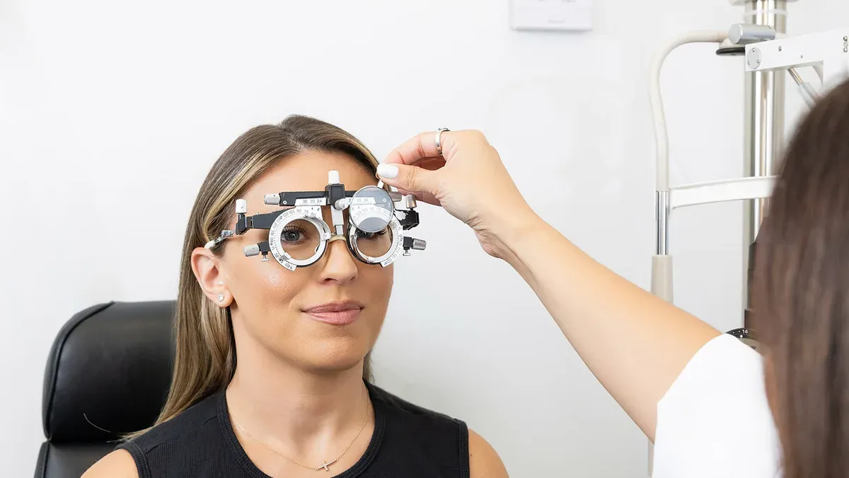
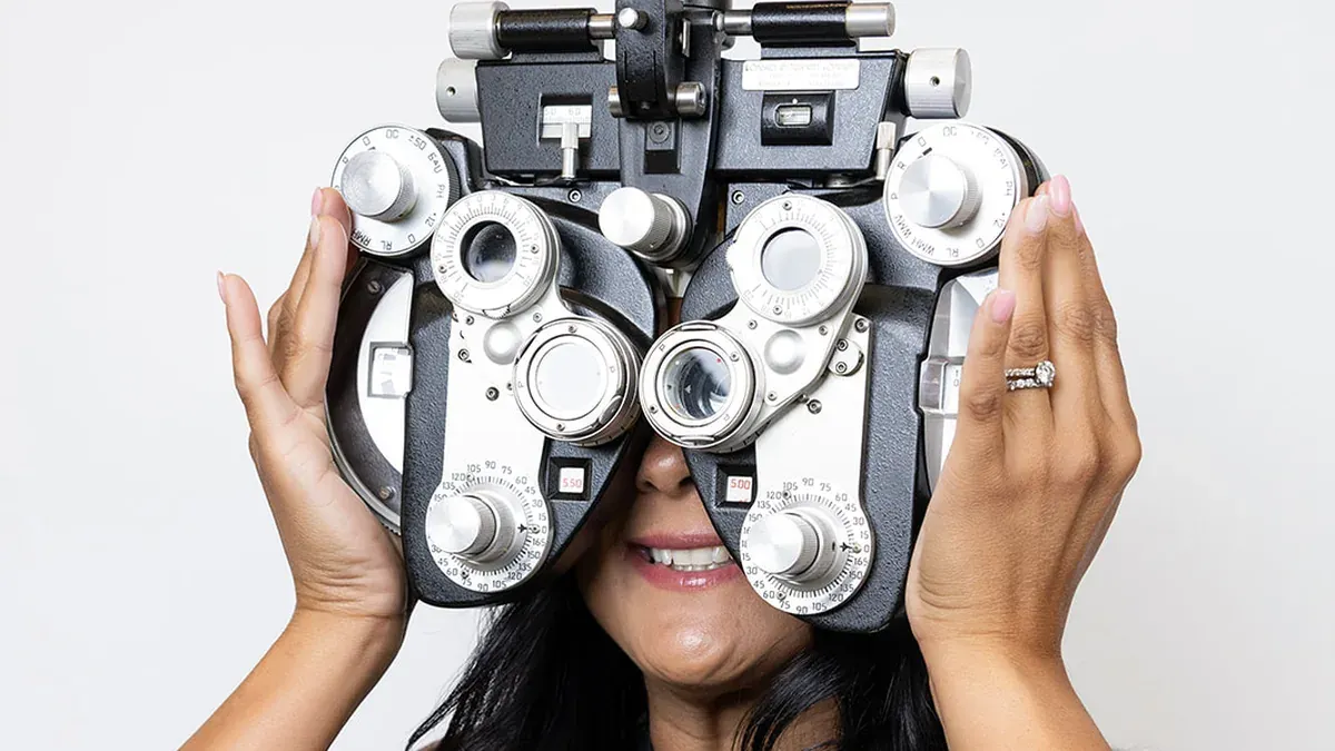
Binocular vision dysfunction (BVD) is a condition in which a subtle misalignment between the eyes forces the brain into constant corrective effort, producing headaches, dizziness, and eye fatigue that bear no relation to how clearly you can read an eye chart. A person can pass every standard acuity test and still live with clinically significant BVD. Because the symptoms closely resemble migraines, anxiety, and vestibular disorders, the condition often goes undiagnosed for years. If chronic symptoms have never been explained by a standard prescription, a comprehensive eye exam that includes binocular vision testing is the right starting point.
What Is Binocular Vision Dysfunction?
Binocular vision dysfunction occurs when the two eyes are slightly out of alignment and the brain cannot receive a stable, fused image without working overtime to compensate. That continuous overcompensation taxes the muscles and neurological pathways involved in visual processing, pushing symptoms well beyond the eyes themselves. A 2025 review by do Amaral Júnior et al. suggested that binocular integration circuits are fine-tuned through extended critical periods in frontal-eyed species.
Signs that the eyes may not be working as a team include:
- Double vision or briefly overlapping images occur when the brain fails to merge both eyes' input, most often during sustained reading or screen use.
- Depth perception errors show up as misjudging curbs, doorframes, or the distance to oncoming objects, particularly in low-contrast or nighttime conditions.
- Near-distance fatigue develops after relatively short sessions of close-up work, often with a pulling sensation behind the eyes.
Why Can You Have 20/20 Vision and Still Have BVD?
Standard acuity tests measure how sharply the eye resolves a target at a fixed distance. They say nothing about the brain's ability to fuse two slightly different images into one stable picture. As Bridge (2016) noted in a review of binocular depth perception, significant cortical processing is required to produce the percept of depth, and brain damage or dysfunction can interfere with stereoscopic vision even when retinal input from both eyes is intact. A patient who reads every chart line correctly can still have binocular misalignment that only surfaces under real-world visual demands: driving at night, prolonged screen work, or moving through a visually complex environment.

BVD Symptoms: Including the Ones That Look Like Something Else
BVD symptoms overlap substantially with migraines, vestibular dysfunction, anxiety disorder, ADHD, and reading disability, routing patients to the wrong specialists for years.
Physical symptoms:
- Tension headaches concentrated at the brow, temples, or base of the skull appear after visually demanding tasks.
- Neck stiffness and shoulder tightness develop as the body compensates for the head position the brain unconsciously adopts to reduce misalignment.
- Facial pressure around the forehead and sinuses occurs without any sinus or allergy cause.
Visual symptoms:
- Blurred vision fluctuates rather than remaining constant, particularly when shifting focus between near and far distances.
- Intermittent double vision or image ghosting appears during reading or screen work, then clears when the person blinks or looks away.
- Light sensitivity in high-glare conditions goes beyond what a standard tint or AR coating resolves.
Reading symptoms:
- Losing place on the line, skipping lines, or re-reading the same line reflects the brain's struggle to track a stable image across the page.
- Comprehension fatigue sets in quickly because working memory carries the extra burden of stabilizing the visual field.
- Early avoidance of reading occurs despite adequate motivation and no diagnosed learning impairment.
Driving symptoms:
- Misjudging the gap between vehicles or the distance to a stop sign reflects disrupted depth perception under binocular conditions.
- Discomfort from oncoming headlights at night goes beyond normal sensitivity because the two eyes process glare asymmetrically.
- Hesitation during lane changes or difficulty judging parking distances involves the same depth-perception mechanism disrupted in BVD.
Anxiety-related symptoms:
- Unease in visually complex or crowded spaces appears because the visual system cannot process competing movement without overload.
- Motion sensitivity on escalators, in moving vehicles, or while scrolling on a phone occurs when visual and proprioceptive signals fail to align.
- Difficulty concentrating in environments with shifting backgrounds or patterned surfaces results from the same processing overload.
Can BVD Cause Headaches, Dizziness, and Anxiety?
Yes. The brain's effort to merge two misaligned images is not passive; it recruits extraocular muscles and neurological pathways continuously, generating the physical tension behind tension headaches and neck pain. Maghlakelidze et al. (2024) noted in a review of binocular interaction that suppression transforms the structurally binocular system into a functionally monocular one, with poor visual functions in amblyopia associated with the presence of suppression in binocular conditions of vision. That same compensatory strain explains why BVD-related dizziness closely mimics vestibular dysfunction, and why the generalized unease some patients experience meets clinical criteria for anxiety disorder.
Can BVD Come on Suddenly?
Yes, BVD can appear abruptly. Concussion, traumatic brain injury, and stroke can disrupt the neurological coordination between the two eyes in a matter of hours, producing acute BVD symptoms where none existed before. A sharp increase in screen demand, such as shifting to a fully remote job, can also push a low-grade lifelong misalignment past the brain's compensation threshold.
Sudden-onset BVD from neurological injury requires evaluation alongside the underlying condition. If new visual symptoms appear with sudden vision loss, severe eye pain, or other acute neurological signs, seek emergency care rather than scheduling a routine appointment.
What Causes Binocular Vision Dysfunction?
Binocular vision dysfunction has several distinct origins, each of which disrupts the brain's ability to maintain a stable, fused image.
- Congenital eye-muscle imbalance produces a structural misalignment present from birth that may remain subclinical until visual demands increase with age or screen use.
- Developmental anomalies in binocular integration circuits affect how visual cortex pathways mature.
- Neurological injury, including concussion, TBI, and stroke, disrupts cortical coordination between the two eyes' signals and can produce BVD onset within days of the event.
- Uncorrected or inadequately corrected refractive error forces one or both eyes to work harder, compounding the brain's fusion burden.
- Age-related changes in eye-muscle coordination reduce the system's tolerance for even small degrees of misalignment that the younger visual system managed without symptoms.

Why a Routine Eye Exam Often Misses BVD
Standard cover tests and acuity charts detect large-angle strabismus and refractive error. BVD sits below that threshold. A person can pass every element of a routine exam and still have clinically significant binocular dysfunction that only surfaces under extended, real-world visual conditions.
The misalignment driving BVD is typically measured in fractions of a prism diopter. Standard clinical tests do not measure at that resolution. At OPTYX, a binocular vision assessment is available as part of a comprehensive eye exam, targeting precisely the degree of misalignment that routine protocols overlook.
How Is BVD Diagnosed?
BVD is diagnosed through a specialized binocular vision evaluation that measures misalignment under real visual conditions, not a standard acuity chart. The scope and measurement precision go well beyond routine testing.
A binocular vision dysfunction test typically includes:
- Detailed symptom history to map the relationship between symptoms and specific visual tasks, screen time, and lighting conditions.
- Binocular vision testing that presents targets to both eyes simultaneously, revealing misalignment that disappears when each eye is tested in isolation.
- Computerized misalignment measurement to quantify the degree and direction of eye misalignment with precision beyond manual prism bars.
- Vergence and accommodation assessment to evaluate how well the eyes converge, diverge, and shift focus under demand.
At OPTYX, the evaluation includes Neurolens technology to quantify eye misalignment precisely and determine candidacy for contoured prism lenses. Standard clinical measurement cannot do this. Neurolens captures misalignment across different gaze angles, giving the optometrist the specific data needed to prescribe a personalized correction.

BVD Treatment: Prism Lenses, Vision Therapy, and Beyond
Once this condition is diagnosed, the primary treatment pathways are prescription prism lenses, structured vision therapy, and, for appropriate candidates, Neurolens contoured prism correction.
BVD treatment options include:
- Prescription prism lenses incorporate a micro-prism into the lens that shifts the light path entering each eye, correcting alignment at the lens so the brain stops overcompensating.
- Structured vision therapy uses guided exercises to retrain binocular coordination over a period of weeks to months.
- Neurolens contoured prism lenses are individually calibrated to the patient's measured misalignment profile and can be built into the patient's chosen frame alongside a standard prescription; candidacy is determined by an OPTYX optometrist based on the full binocular evaluation.
- Combination treatment, using prism correction alongside vision therapy, is prescribed when the degree of misalignment warrants both structural correction and muscle retraining.
Neurolens is designed to relieve symptoms linked to eye misalignment. It is not described as a cure or a treatment of an underlying structural cause.
What Do BVD Glasses (Prism Lenses) Look Like?
Prism lenses look identical to ordinary prescription glasses. The prism correction is ground into the lens material itself, leaving no visible wedge or additional element. Someone wearing BVD glasses in a standard frame could not be identified as such by an observer.
Adaptation after a new prism prescription typically takes a few days to a few weeks, depending on the degree of correction. Some patients notice a mild sense of spatial shift in the first days as the visual system adjusts to properly aligned input. An OPTYX optometrist monitors adaptation and can refine the prescription as needed.
When to Get Evaluated for BVD
If you have experienced persistent headaches, dizziness, neck tension, reading difficulty, or unease in visually busy environments that has not been explained by a standard prescription, binocular vision dysfunction is worth evaluating. Standard exams are not built to find these symptoms.
OPTYX offers specialized binocular vision assessment as part of a comprehensive eye exam, available across OPTYX locations in Manhattan, Long Island, the North Fork, the Hamptons, and Westchester. Book an Eye Exam and ask about binocular vision testing at the time of booking.
Sources
- do Amaral Júnior FL et al. (2025). Comparative Neuroplasticity in Frontal- and Lateral-Eyed Mammals With Induced-Binocular Vision Dysfunction: Insights From Monocular Deprivation Models. European Journal of Neuroscience. PubMed
- Maghlakelidze N et al. (2024). Binocular Interaction in Amblyopia. Georgian Medical News. PubMed
- Bridge H (2016). Effects of Cortical Damage on Binocular Depth Perception. Philosophical Transactions of the Royal Society of London, Series B, Biological Sciences. PubMed