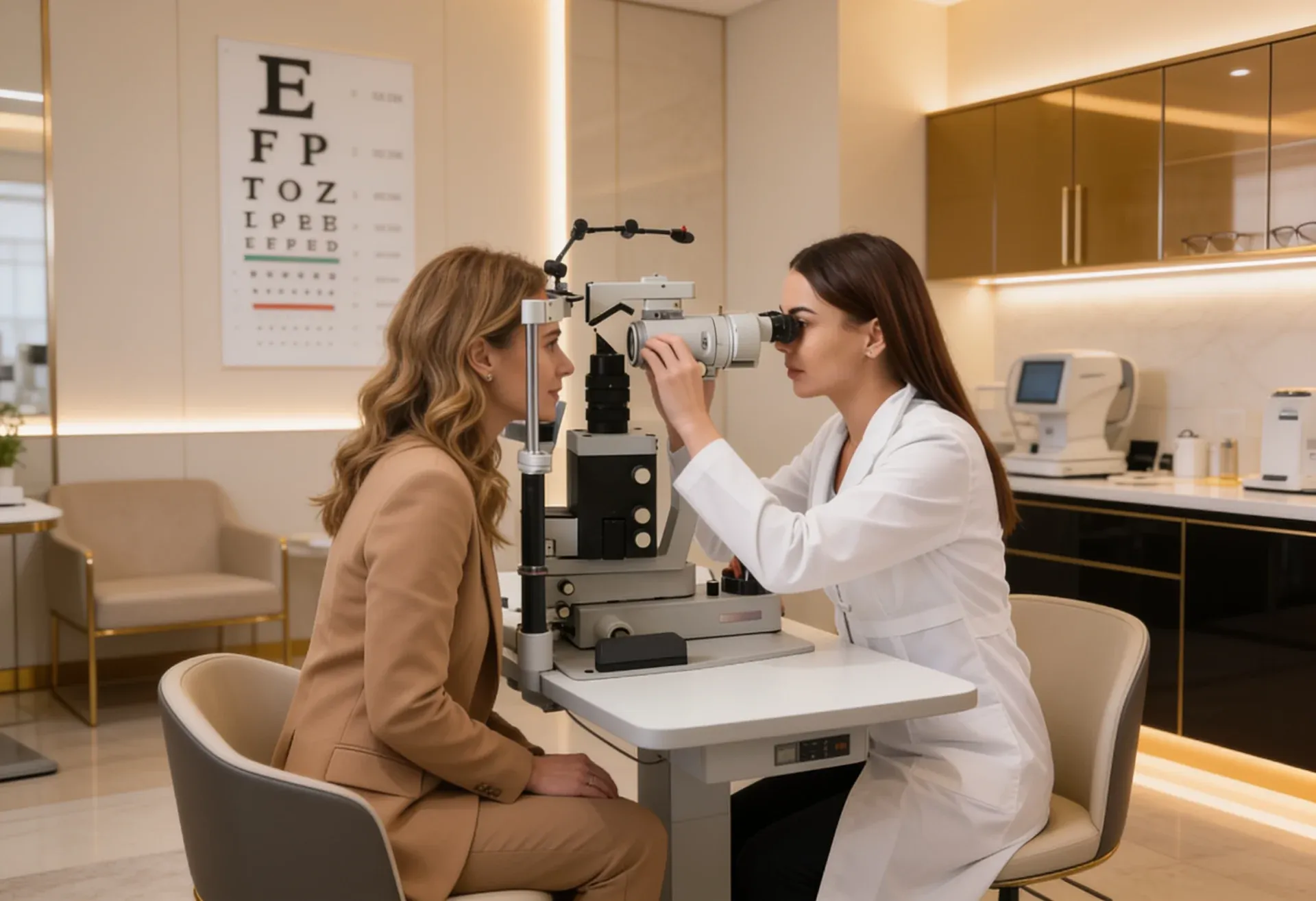

In the sophisticated realm of contemporary eye care, where technological precision meets clinical excellence, few conditions challenge diagnostic capabilities quite like atrophic retinal holes. These subtle peripheral retinal breaks, often measuring mere millimeters in diameter, represent a fascinating intersection of degenerative pathology and cutting-edge detection technology. At OPTYX, the premier destination for advanced eye care since 1971, we’ve elevated the detection of these elusive lesions to an art form through our mastery of Optos® ultra-widefield imaging.
The landscape of retinal diagnostics has evolved dramatically over the past decade, with atrophic hole optos® technology leading this transformation. Unlike the dramatic presentations of acute retinal tears or detachments, atrophic retinal holes whisper their presence through subtle morphological changes that demand the most sophisticated detection methods available.
The Anatomy of Atrophic Retinal Holes: Understanding the Basics
Atrophic retinal holes are small breaks that develop in the outer edges of the retina – the light-sensitive tissue at the back of your eye. Think of them as tiny holes that form naturally over time, usually in the peripheral areas where your central vision doesn’t rely on.
These holes develop differently from other retinal problems. Instead of being caused by sudden injury or pulling forces, atrophic holes form gradually as the retinal tissue slowly thins out with age or other natural changes. They’re typically round or oval-shaped, ranging from very small (almost microscopic) to a few millimeters across.
What makes these holes particularly challenging to detect is their location. They usually appear in the far edges of the retina, well beyond what traditional eye examination methods can see clearly. This is where the advanced atrophic retinal hole optos® imaging at OPTYX becomes essential – it can capture these remote areas with exceptional detail.
The key difference between atrophic holes and other retinal breaks is how they form. While some retinal tears happen when the vitreous gel pulls on the retina, atrophic holes develop when the retinal tissue itself gradually weakens and creates a natural opening. This makes them quieter and more subtle, often going unnoticed without advanced imaging technology.

The Technical Superiority of Optos® Technology in Atrophic Hole Detection
The detection of atrophic retinal holes demands imaging technology that can capture the peripheral retina with exceptional clarity and detail. Optos® ultra-widefield imaging systems achieve this through sophisticated scanning laser ophthalmoscopy, employing multiple wavelengths to penetrate retinal layers and create comprehensive maps of retinal architecture.
The technical specifications of atrophic hole optos® imaging at OPTYX include capture of up to 200 degrees of retinal detail in a single acquisition, compared to the limited 30-45 degree field of view provided by traditional fundus photography. This expanded field of view is crucial for atrophic hole detection, as these lesions frequently occur in the far periphery where conventional imaging methods cannot reach.
Our atrophic retinal hole optos® protocol employs both red and green laser wavelengths, each optimized for different aspects of retinal visualization. The red laser provides excellent visualization of deeper retinal structures, while the green laser offers superior contrast for retinal surface features and vascular patterns. This dual-wavelength approach ensures that atrophic retinal hole on optos® images captures both the morphological characteristics of the holes themselves and the surrounding retinal context essential for accurate diagnosis.
Advanced Image Analysis and Interpretation Techniques
The sophistication of modern atrophic hole optos® technology extends beyond image acquisition to encompass advanced analysis and interpretation capabilities. At OPTYX, our eye care professionals utilize specialized software tools that enhance image contrast, adjust color balance, and provide measurement capabilities essential for accurate lesion characterization.
The interpretation of atrophic retinal hole optos® images requires an understanding of the subtle morphological features that distinguish these lesions from other peripheral retinal abnormalities. True atrophic holes appear as well-demarcated, round or oval defects with smooth edges, often surrounded by areas of retinal pigment epithelium changes or associated lattice degeneration.
Digital enhancement capabilities allow our optometrists to optimize atrophic retinal hole on optos® images for maximum diagnostic value. These tools include contrast adjustment, color mapping, and magnification features that reveal details invisible to conventional examination methods. The ability to manipulate image parameters in real-time during patient consultation enhances both diagnostic accuracy and patient education.
Quality Assurance and Technical Standards
The technical excellence of atrophic retinal hole optos® imaging at OPTYX is maintained through rigorous quality assurance protocols and ongoing technical training. Our imaging systems undergo regular calibration and maintenance to ensure optimal performance, while our eye care professionals participate in continuous education programs focused on advanced imaging interpretation.
Image quality standards for atrophic hole optos® documentation include assessment of focus, illumination uniformity, and artifact minimization. Each atrophic retinal hole on optos® image is evaluated for technical adequacy before being incorporated into the patient’s permanent record, ensuring that diagnostic decisions are based on the highest quality imaging data available.
Key Advantages of Our Detection Protocol:
- Ultra-widefield imaging capturing up to 200 degrees of retinal detail
- High-resolution visualization of peripheral retinal structures
- Non-invasive scanning with a comfortable patient experience
- Immediate image availability for real-time consultation
- Advanced digital enhancement capabilities for optimal diagnosis
- Comprehensive documentation for long-term monitoring
- Integration with complete eye examination protocols
- Expert interpretation by experienced eye care professionals
Frequently Asked Questions
What makes atrophic retinal hole optos® imaging superior to traditional methods?
The ultra-widefield capability captures peripheral retinal areas invisible to conventional fundoscopy, while high-resolution imaging reveals subtle morphological details essential for accurate diagnosis.
How often should atrophic hole optos® imaging be performed?
Imaging frequency depends on individual risk factors and the presence of existing lesions. Our optometrists develop personalized monitoring schedules based on comprehensive risk assessment.
Can an atrophic retinal hole on optos® imaging detect other peripheral pathology?
Absolutely. The comprehensive peripheral visualization reveals lattice degeneration, peripheral tears, retinoschisis, and various other conditions that might otherwise go undetected.
What technical factors affect atrophic retinal hole optos® image quality?
Key factors include patient cooperation, media clarity, pupil size, and proper device calibration. Our technical protocols optimize these variables for maximum image quality.
How does OPTYX ensure consistent atrophic hole optos® imaging quality?
Through rigorous equipment maintenance, ongoing staff training, standardized protocols, and comprehensive quality assurance programs that maintain the highest technical standards.
Take Action: Experience Technical Excellence in Eye Care
Don’t compromise on the technical quality of your eye care. Schedule your comprehensive eye examination with advanced atrophic retinal hole optos® imaging at OPTYX today and experience the difference that cutting-edge technology and technical expertise make in preserving your vision.
Contact OPTYX at our convenient New York City or Long Island locations to learn more about our advanced imaging capabilities and comprehensive approach to retinal health monitoring. Your vision deserves the technical excellence that only OPTYX can provide.
Visit OPTYX today – where technical innovation meets clinical excellence in the pursuit of optimal eye health.