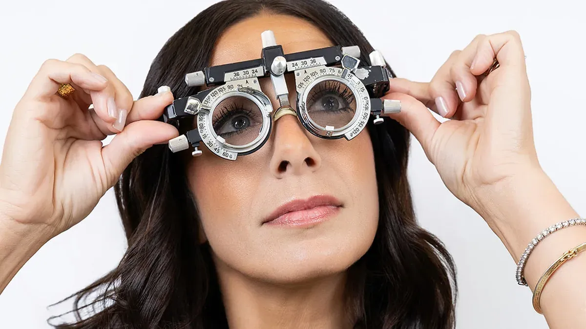

Eye exam equipment is the collection of vision testing instruments your optometrist uses to measure how clearly you see and how healthy your eyes are. Some are simple to describe. Others, like the tonometer that measures internal eye pressure, detect risk factors for conditions such as glaucoma that cause no symptoms on their own. Every instrument earns its place.
What Tools Does an Eye Doctor Use During an Exam?

A complete exam draws on several categories of eye exam equipment, each designed for a different layer of vision or ocular health. The table below maps the core instruments to what they measure.
| Tool | What It Measures |
|---|---|
| Snellen chart | Central visual acuity at distance |
| Phoropter | Refractive error and lens prescription |
| Autorefractor | Automated refractive error estimate |
| Slit lamp biomicroscope | Cornea, iris, lens, and anterior segment |
| Applanation tonometer | Intraocular pressure (IOP) |
| Non-contact tonometer | IOP via brief air puff |
| Retinal imaging camera | Retina, optic nerve, and macula |
| Visual field perimeter | Peripheral vision and blind spots |
The table is a quick reference. Each section below explains what the instrument does and why your optometrist uses it at that stage of the exam.
What Equipment Is Used to Test Visual Acuity?
The Snellen chart is the most recognizable piece of eye exam equipment in any practice. It measures Snellen chart visual acuity by presenting rows of letters that shrink with each line, read from 20 feet. A result of 20/20 means you can read at 20 feet what someone with typical vision also reads at that distance. Numbers tell the story.
Patients who cannot read letters use the tumbling-E chart, which presents the same letter in four different orientations. Near vision is tested with the Jaeger chart, a card printed with text ranging from large to fine print, to establish whether reading glasses may be needed. Learn how Snellen chart visual acuity testing converts those readings into a prescription number.
What Is a Phoropter Used for in Vision Testing?

The phoropter is the mask-like instrument your optometrist uses during the refraction exam, housing a rotating drum of precisely ground lenses. You respond yes or no as lens pairs switch during the phoropter vision test, and those responses narrow down your refractive error to produce a final prescription.
Before the phoropter phase, most practices run an autorefractor measurement first. At OPTYX, an autorefractor shines a small infrared beam into the eye, measures how it reflects off the retina, and generates an automated refractive estimate in seconds. That reading gives the optometrist a starting baseline before the phoropter dialogue begins. Your responses shape everything.
What Is a Slit Lamp Used for During an Eye Exam?
The slit lamp biomicroscope lets your optometrist examine the front of the eye under high magnification. A narrow, intensely focused beam illuminates the cornea, iris, anterior chamber, and crystalline lens in fine detail, making early cataract changes, surface damage, and signs of inflammation visible in a single pass. Nothing escapes that beam.
No other piece of eye exam equipment delivers the same combined view of the anterior segment. The slit lamp is also the primary diagnostic tool for detecting dry eye and irritation at the ocular surface, where changes in the tear film first appear. Angling the beam lets the optometrist isolate individual tissue layers.
How Does a Tonometer Measure Eye Pressure?

A tonometer measures intraocular pressure (IOP) by assessing how the cornea responds to contact. Pressure rarely shows symptoms. The applanation tonometer is the clinical gold standard: after a numbing drop, a small flat probe briefly flattens a defined area of the cornea, and the force required corresponds directly to the pressure inside the eye.
The alternative is the non-contact tonometer. It measures IOP by directing a brief burst of air at the cornea without any physical contact, making it common in routine screenings. For detailed glaucoma monitoring, the applanation tonometer is preferred. Find out more about how the air-puff tonometry test works and what it checks for.
Retinal Imaging Camera: Key Eye Exam Equipment for Early Detection
A retinal imaging camera photographs the retina, optic nerve, and blood vessels at the back of the eye. Those images let your optometrist monitor for diabetic retinopathy, age-related macular degeneration, and changes to the optic nerve head over time, often before any symptoms appear. That changes everything.
Standard retinal cameras capture a central field around the optic nerve and macula. Wider-field systems like Optos can image up to 200 degrees of the retina in a single frame, often without dilation, reaching peripheral areas that narrower cameras miss. Retinal imaging has become a routine part of a comprehensive exam, not a specialist-only procedure.
Book an Eye Exam
To see this eye exam equipment matched to your specific vision and health picture, Book an Eye Exam at any OPTYX New York location.