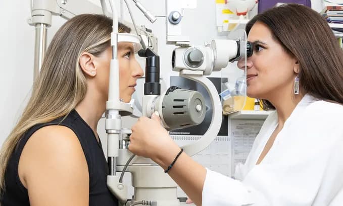

The field of retinal imaging has been revolutionized by the introduction of advanced Optos® fundus photo technology, which represents a significant leap forward from traditional fundus photography methods. This innovative imaging approach has transformed how eye care professionals diagnose, monitor, and treat various retinal conditions, offering unprecedented visualization capabilities that were previously impossible with conventional techniques.
What is Optos® Fundus Photo Technology
Optos® fundus photo technology utilizes ultra-widefield scanning laser ophthalmoscopy to capture comprehensive retinal images that extend far beyond the capabilities of traditional fundus photography. Unlike conventional methods that typically capture only 30-45 degrees of the retina, Optos® fundus photo systems can image up to 200 degrees of the retinal surface in a single capture, providing a panoramic view of the eye’s interior structures.
At OPTYX New York, our optometry practice utilizes state-of-the-art Optos® imaging systems that employ multiple laser wavelengths to create detailed, high-resolution images of the retina. This advanced Optos® photo technology enables our optometrists to detect peripheral retinal pathology that might otherwise go unnoticed during standard examinations.

Optos® Fundus Photo Image Acquisition Process
The Optos® fundus photo process is remarkably efficient, typically requiring only seconds to capture comprehensive retinal images. The system’s ability to image through undilated pupils represents a significant advantage over traditional fundus photography, which often requires time-consuming pupil dilation and can cause patient discomfort.
During an Optos® fundus photo examination at our optometry practice, patients simply look into the device while the system automatically captures ultra-widefield images of their retina. The process is quick, comfortable, and provides immediate results that can be reviewed with the patient during their appointment.
Optos® vs Fundus Photo: Comprehensive Comparison
When comparing Optos® vs fundus photo methodologies, several critical differences emerge that highlight the advantages of ultra-widefield imaging technology. Traditional fundus photography has served as the standard for retinal documentation for decades, but it comes with inherent limitations that Optos technology addresses.
Field of View
The most significant difference in Optos® vs fundus photo comparison lies in the field of view captured. Traditional fundus photography typically captures 30-45 degrees of the retina, focusing primarily on the posterior pole, including the macula and optic disc. In contrast, Optos® fundus photo technology captures up to 200 degrees, providing visualization of peripheral retinal areas that are crucial for comprehensive eye health assessment.
This expanded field of view in Optos® vs fundus photo comparison means that conditions such as peripheral retinal tears, diabetic retinopathy changes, and retinal detachments can be detected earlier and more comprehensively. At OPTYX New York, this capability has significantly enhanced our diagnostic accuracy and treatment planning in our optometric practice.
Patient Experience: Optos® vs Fundus Photo
Another important aspect of the Optos® vs fundus photo comparison involves patient experience. Traditional fundus photography often requires pupil dilation, which can cause light sensitivity and blurred vision for several hours following the examination. Patients may need to arrange transportation and take time off work due to these side effects.
Optos® fundus photo technology eliminates most of these concerns by imaging through undilated pupils in the majority of cases. This advancement makes comprehensive retinal examinations more convenient and comfortable for patients, encouraging better compliance with regular optometric care appointments.
Image Quality in Optos® Fundus Photo Systems
The Optos® vs fundus photo comparison also reveals significant differences in image quality and diagnostic capability. While traditional fundus photography provides excellent detail of the central retina, Optos® fundus photo technology offers superior visualization of peripheral retinal structures while maintaining high-resolution imaging of central areas.
Multi-Modal Optos® Fundus Photo Capabilities
The multi-modal capabilities of Optos® fundus photo systems allow for various imaging techniques, including color photography, autofluorescence, and fluorescein angiography, all within the ultra-widefield format. This versatility provides comprehensive diagnostic information that enhances decision-making in our optometry practice.
Optos fundus photo systems can capture different types of images during a single examination session, providing multiple perspectives of retinal health. This comprehensive approach ensures that subtle pathological changes are not missed during routine optometric examinations.
Advantages of Optos® Fundus Photo Technology
At OPTYX New York, our investment in advanced optos fundus photo technology reflects our commitment to providing the highest standard of optometric care. The comprehensive imaging capabilities allow our optometrists to make more accurate diagnoses and develop more effective treatment plans.
Enhanced Diagnostic Accuracy with Optos® Fundus Photo
The superior field of view provided by Optos® fundus photo technology enables the detection of retinal pathology that might be missed with traditional methods. This enhanced diagnostic capability leads to earlier intervention and better patient outcomes in our optometry practice.
The detailed documentation provided by Optos® fundus photo systems allows for precise monitoring of disease progression and treatment effectiveness over time.
Improved Patient Experience
The convenience and comfort of Optos® photo examinations encourage patients to maintain regular optometric care appointments. The quick, non-invasive nature of the imaging process makes comprehensive retinal evaluations more accessible to patients with busy schedules or those who are sensitive to bright lights.
Comprehensive Documentation
Optos® fundus photo technology provides detailed documentation of retinal health that can be compared over time to monitor disease progression or treatment effectiveness. This longitudinal tracking capability is essential for managing chronic retinal conditions and provides valuable information for treatment planning in our optometric practice. format. This versatility provides comprehensive diagnostic information that enhances decision-making in our optometry practice.
Conclusion
The comparison of Optos® vs fundus photo clearly demonstrates the superior capabilities of ultra-widefield imaging technology. Optos fundus photo systems provide comprehensive retinal visualization, enhanced patient comfort, and improved diagnostic accuracy compared to traditional fundus photography methods.
At OPTYX New York, our optometry practice is committed to utilizing the most advanced Optos® fundus photo technology to provide our patients with exceptional eye care. The comprehensive imaging capabilities of our Optos systems ensure that our patients receive the most thorough retinal evaluations possible from our experienced optometrists.
If you would like to experience the benefits of ultra-widefield retinal imaging through Optos® fundus photo technology, we invite you to schedule a comprehensive eye examination with our experienced optometry team. Visit OPTYX New York to learn more about our advanced imaging services and book your appointment today.