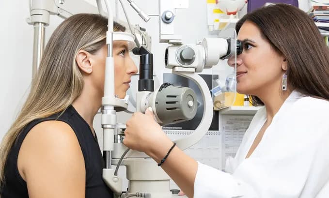

The diagnosis and management of retinoschisis have been significantly enhanced through the implementation of advanced retinoschisis Optos® imaging technology. This sophisticated ultra-widefield imaging approach has revolutionized how optometrists detect, monitor, and differentiate retinoschisis from other retinal conditions, providing unprecedented visualization capabilities that are essential for accurate diagnosis and effective patient care.
Understanding Retinoschisis and Optos® Technology
Retinoschisis is a condition characterized by the splitting of the retinal layers, creating a separation within the neurosensory retina itself. Unlike retinal detachment, where the entire neurosensory retina separates from the retinal pigment epithelium, retinoschisis involves an intraretinal split that can be challenging to detect with traditional imaging methods.
At OPTYX New York, our optometry practice utilizes state-of-the-art retinoschisis Optos® systems that provide comprehensive ultra-widefield imaging capabilities. This advanced technology enables our optometrists to visualize the entire retinal surface, including peripheral areas where retinoschisis commonly occurs, ensuring accurate diagnosis and appropriate treatment planning.

Optos® Technology for Retinoschisis Detection
The implementation of Optos® retinoschisis imaging has transformed the diagnostic approach to retinal layer separation conditions. Optos® ultra-widefield systems capture up to 200 degrees of the retinal surface in a single image, providing comprehensive visualization that extends far beyond the capabilities of traditional fundus photography.
Advanced Imaging Capabilities
Optos® retinoschisis detection utilizes scanning laser ophthalmoscopy combined with multiple imaging modalities to provide detailed visualization of retinal layer architecture. The system’s ability to capture high-resolution images without pupil dilation in most cases makes the examination process more comfortable for patients while providing superior diagnostic information.
The multi-modal capabilities of retinoschisis Optos® systems include color photography, autofluorescence imaging, and fluorescein angiography, all within the ultra-widefield format. This comprehensive imaging approach enables optometrists to assess the extent and characteristics of retinoschisis with exceptional detail and accuracy.
Retinoschisis vs Retinal Detachment Optos®: Critical Diagnostic Differences
One of the most important applications of advanced imaging technology involves the differentiation between retinoschisis and retinal detachment. The comparison of retinoschisis vs retinal detachment Optos® imaging reveals critical diagnostic features that are essential for appropriate treatment planning and patient management.
Distinguishing Features on Optos Imaging
When examining retinoschisis vs retinal detachment Optos® images, several key characteristics help differentiate these conditions. Retinoschisis typically appears as a smooth, dome-shaped elevation with a characteristic appearance that differs significantly from the irregular, mobile nature of retinal detachment.
The ultra-widefield capabilities of retinoschisis vs retinal detachment Optos® imaging allow optometrists to visualize the full extent of retinal abnormalities, including peripheral areas that might be missed with conventional imaging methods. This comprehensive view is particularly important for distinguishing between these two conditions, as the extent and pattern of involvement can provide crucial diagnostic information.

Benefits of Optos® Technology at OPTYX New York
At OPTYX New York, our investment in advanced retinoschisis optos technology reflects our commitment to providing the highest standard of optometric care. The comprehensive imaging capabilities enable our optometrists to deliver more accurate diagnoses and develop more effective treatment plans for patients with retinal conditions.
Enhanced Diagnostic Precision
The superior imaging capabilities of our Optos® systems enable the detection of retinoschisis and other retinal conditions with exceptional accuracy. This enhanced diagnostic precision leads to earlier intervention when necessary and better patient outcomes overall.
Comprehensive Patient Care
The detailed documentation provided by Retinoschisis Optos® Imaging supports comprehensive patient care by enabling precise monitoring of condition progression and treatment effectiveness over time. This longitudinal tracking capability is essential for optimal patient management.
Professional Excellence
Our optometrists utilize the latest advances in retinoschisis Optos® technology to ensure patients receive the most advanced diagnostic capabilities available. This commitment to technological excellence supports our mission of providing exceptional eye care services.
Conclusion
The implementation of advanced retinoschisis optos® imaging technology has revolutionized the diagnosis and management of retinal layer separation conditions. The superior imaging capabilities, enhanced diagnostic accuracy, and improved patient experience provided by this technology make it an essential tool for comprehensive optometric care.
At OPTYX New York, our optometry practice is committed to utilizing the most advanced retinoschisis Optos® technology to provide our patients with exceptional diagnostic capabilities and treatment outcomes. The comprehensive imaging capabilities of our Optos® systems ensure that patients receive the most thorough retinal evaluations possible from our experienced optometry team.
If you are concerned about retinal health or would like to experience the benefits of advanced ultra-widefield retinal imaging, we invite you to schedule a comprehensive eye examination with our experienced optometrists. Visit OPTYX to learn more about our advanced imaging services and book your appointment today.