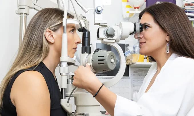

When you think about an eye exam, you probably picture the letter chart on the wall. But a visual field eye exam tests something completely different. Not how sharp your central vision is. How much you can see. At OPTYX, a visual field test eye exam maps the full range of your peripheral and central vision to find gaps you cannot feel and cannot notice, because your brain quietly fills them in.
What Is a Visual Field Eye Exam?
What is a visual field eye exam exactly? It is a test that maps your entire field of vision, from the central point you focus on to the outer edges of your peripheral sight. It measures sensitivity at hundreds of points across your visual field, not just whether you can read a letter at a given size.
Understanding what is a visual field eye exam helps explain why it catches conditions that a standard vision test misses entirely. Many serious diseases damage peripheral vision first, gradually enough that you adapt without noticing. This assessment catches those changes before they affect your daily life.
What Does an Eye Exam Visual Field Test Measure?
An eye exam visual field test measures the sensitivity of your retina and optic nerve at points across your whole visual field. In a standard automated test, you sit in front of a curved screen, keep your gaze on a central fixation point, and press a button each time you see a flash of light. The flashes vary in position and brightness.
The eye exam visual field test produces a grayscale sensitivity map compared against normal values for your age. Your optometrist looks for:
- Scotomas: areas where sensitivity is reduced or gone entirely. You would not notice these in daily life because your brain fills in the blank.
- Peripheral field loss: narrowing at the edges of your vision, one of the hallmarks of glaucoma.
- Central field loss: gaps near the center, common in macular disease.
- Hemianopia: loss of half the visual field, which can point to stroke or a neurological condition.
Why Does Visual Field Testing Matter?
This assessment is so important because the conditions it catches produce no early symptoms. Glaucoma is the clearest example. It damages the optic nerve starting at the peripheral edges of your vision, so gradually that most patients do not notice until significant damage has already occurred.
A visual field test eye exam detects those peripheral gaps early, when treatment can still slow or stop further damage. Beyond glaucoma, the test also monitors:
- Diabetic retinopathy affecting peripheral sensitivity before central vision changes.
- Optic neuritis and optic neuropathy, where defects follow specific nerve patterns.
- Neurological conditions including stroke, multiple sclerosis, and brain tumors, each producing characteristic patterns on the map.
- Medication toxicity, including hydroxychloroquine (Plaquenil), which can silently damage the macula over time.
What to Expect During the Test
A visual field test eye exam takes 10 to 15 minutes per eye:
- You rest your head against a chin and forehead support, with one eye covered at a time.
- Keep your gaze on the central fixation point throughout. Moving your eyes affects the accuracy of the results.
- Press the button each time you see a flash, even a very faint one.
- Some points are tested more than once. Variability between visits is normal, and a single result rarely determines a diagnosis on its own.

How It Connects to Glaucoma
Glaucoma is the most common reason this test is ordered. The optic nerve damage creates characteristic arc-shaped defects in the peripheral visual field, starting in the nasal region before spreading inward.
Serial results from an eye exam visual field test, checked every six to twelve months, show whether the disease is stable or progressing. At OPTYX, the assessment is paired with tonometry and retinal imaging to build a complete picture of optic nerve health. Read more about how glaucoma pressure testing works alongside visual field testing.
How Often Do You Need It?
For patients without known risk factors, baseline visual field testing from age 40 onward is standard. For patients with glaucoma or elevated eye pressure, the test every six months is typical.
If you take medications known to affect vision, have a neurological condition, or have a first-degree relative with glaucoma, your optometrist may recommend more frequent monitoring.
Book a Comprehensive Eye Exam at OPTYX
The eye exam visual field test is included in every comprehensive exam at OPTYX. Our optometrists pair it with retinal imaging and tonometry so nothing gets missed.
OPTYX has 14 locations across Manhattan, Long Island, the North Fork, the Hamptons, and Westchester. Book your eye exam and include visual field testing in your assessment.
Frequently Asked Questions
What is a visual field eye exam?
This test maps the full extent of your peripheral and central vision, detecting blind spots or reduced sensitivity you would never notice on your own. It is most commonly used to screen for and monitor glaucoma, but also identifies neurological conditions and other diseases affecting the visual pathway.
What does a visual field test eye exam involve?
You sit in front of a curved screen, keep your gaze on a central fixation point, and press a button each time you see a flash of light. The test takes 10 to 15 minutes per eye and produces a sensitivity map compared against normal values for your age.
How does an eye exam visual field test detect glaucoma?
Glaucoma causes progressive optic nerve damage starting in your peripheral visual field. The test detects those peripheral gaps early, sometimes years before you notice any change, when treatment can still prevent further damage.
Does the visual field test hurt?
No. It is completely non-invasive. You sit and respond to light flashes. No drops, no contact with your eye, no discomfort.
How often should I have a visual field eye exam?
If you have glaucoma or elevated eye pressure, testing every six months is typically recommended. For other patients, annual testing from age 40 onward is standard practice.
What happens if my result shows an abnormality?
Your optometrist will look at the pattern of the defect, compare it with previous results if available, and determine whether further testing or a specialist referral is needed. A single abnormal result does not automatically mean a diagnosis.